鼻中隔軟骨
外觀
| 鼻中隔軟骨 | |
|---|---|
 鼻子的骨頭和藍色的鼻中隔軟骨 | |
 從下方觀察的鼻軟骨與藍色的鼻中隔軟骨 | |
| 標識字符 | |
| 拉丁文 | Cartilago septi nasi |
| TA98 | A06.1.01.013 |
| TA2 | 946 |
| FMA | FMA:59503 |
| 格雷氏 | p.992 |
| 《解剖學術語》 [在維基數據上編輯] | |
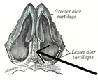
鼻中隔軟骨(英語:septal nasal cartilage)是由透明軟骨所組成[1]。某些地方看起來像四邊形,其邊緣比中間還要厚實。鼻中隔軟骨把前面鼻腔的中間部分給分成左右兩邊,形成兩個鼻孔。
鼻中隔軟骨的前緣之上連接著鼻骨一直到鼻翼小軟骨的前緣;前緣之下連接到鼻翼大軟骨的內側纖維組織。
鼻中隔軟骨的後緣連接著篩骨垂直板;其下緣與犁骨及上頜骨的齶突連在一起。
參見
[編輯]參考資料
[編輯]本條目包含來自屬於公共領域版本的《格雷氏解剖學》之內容,而其中有些資訊可能已經過時。
- ^ Saladin, Kenneth S. Anatomy and Physiology 6th. New York, NY: McGraw Hill, 2012.: McGraw Hill Higher Education. : 856. ISBN 9780077472139.
外部連結
[編輯]- 圖譜:rsa1p7 at the University of Michigan Health System - "Nasal septum, lateral view"
- Anatomy figure: 33:02-01 at Human Anatomy Online, SUNY Downstate Medical Center - "Diagram of skeleton of medial (septal) nasal wall."
- (英文)lesson9 在韋斯利諾曼的解剖課上(喬治城大學) (nasalseptumbonescarti)
