椎动脉
| 椎动脉 | |
|---|---|
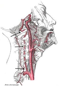
 颈部的动脉:椎动脉起源自锁骨下静脉,最终汇合成 基底动脉 | |
| 基本信息 | |
| 来源 | 锁骨下动脉 |
| 分支 | 基底动脉 脊髓后动脉 脊髓前动脉 小脑后下动脉 |
| 静脉 | 椎静脉 |
| 标识字符 | |
| 拉丁文 | arteria vertebralis |
| MeSH | D014711 |
| TA98 | A12.2.08.002 |
| TA2 | 4538 |
| FMA | FMA:3956 |
| 格雷氏 | p.578 |
| 《解剖学术语》 [在维基数据上编辑] | |
椎动脉(vertebral artery)是颈部的主要动脉之一。一般情况下,椎动脉起自两侧的锁骨下动脉,左右各一,向上穿过第6~1颈椎横突孔,经枕骨大孔入脑汇合成基底动脉。 椎动脉向脊髓上部、脑干、小脑和大脑后部供血。[1]
结构
[编辑]椎动脉通常起源于两侧锁骨下静脉的后上方[2],之后进入第六颈椎(C6)的横突孔,少数情况下(7.5%)进入隆椎(C7)的横突孔。[1]之后,椎动面在各级颈椎的横突孔内上行,穿过寰椎(C1)的横突孔后,跨(穿)寰椎后弓(有椎动脉压迹或弓形孔),经枕骨大孔进入颅内。[1]在颅内、脑桥的底部,两条椎动脉汇合形成基底动脉。在每一级颈椎,椎动脉发出脊柱前动脉分支营养周围的肌肉组织。
变异
[编辑]椎动脉的走向和大小多变,两侧的椎动脉直径常不对称。[3] 例如,左右椎动脉大小的差异可能从轻微不对称到一侧明显发育不全不等。研究估计,单侧椎动脉发育不全的流行率在2%-25%之间。[4]在3-15%的人群中,名为弓形孔的骨桥覆盖了寰椎(C1)上的椎动脉压迹。很少有椎动脉由C1-C2(3%)或C2-C3(仅报道了三例)椎骨水平而不是寰枕水平进入蛛网膜下腔。[5]
椎动脉优势
[编辑]椎动脉(Vertebral artery dominance, VAD) 是一种先天的正常的血管变异,两侧的椎动脉直径将会存在差异,直径较大的一侧为主导侧,直径较小的一侧为非主导侧。有研究表明,约有54%的人为左侧椎动脉优势,30%为右侧椎动脉优势,16%两侧椎动脉直径相近。[6]
诊断
[编辑]
通常使用多普勒超声、断层血管摄影、PC-MRI评估颈动脉的健康状态。
椎颈动脉血液流速的经典评价指标是收缩期峰值流速(PSV)和舒张末期流速(EDV)。[7]
正常情况下, 根据Kuhl等人的一项研究,椎动脉的PSV期血液流速为63.6 ± 17.5 cm/s,EDV期血液流速为16.1 ± 5.1 cm/s。[8] 由于椎动脉优势, 两侧的测量结果可能存在差异。 例如, Seidel 等人进行的另一项研究发现,在PSV期,右侧椎动脉平均流速为45.9 cm/s,而左侧椎动脉的平均流速为51.5 cm/s;在EDV期右侧椎动脉平均流速为13.8 cm/s,而左侧椎动脉的平均流速为16.1 cm/s。[7][9]
其它图像
[编辑]-
大脑底部的动脉(底面观)。
-
脑底部动脉循环图。
-
椎动脉与枕下肌的关系。
参考文献
[编辑]- ^ 1.0 1.1 1.2 Standing S, Borely NR, Collins P, Crossman AR, Gatzoulis MA, Healy GC, et al. Gray's Anatomy: The Anatomical Basis of Clinical Practice 40th. London: Churchill Livingstone. 2008. ISBN 978-0-8089-2371-8.
- ^ Yuan SM. Aberrant Origin of Vertebral Artery and its Clinical Implications. Brazilian Journal of Cardiovascular Surgery. February 2016, 31 (1): 52–9. PMC 5062690
 . PMID 27074275. doi:10.5935/1678-9741.20150071.
- ^ Sun, Yan; Shi, Yan-Min; Xu, Ping. The Clinical Research Progress of Vertebral Artery Dominance and Posterior Circulation Ischemic Stroke. Cerebrovascular Diseases. 2022-02-03, 51 (5): 553–556 [2024-03-09]. ISSN 1015-9770. doi:10.1159/000521616. (原始内容存档于2024-03-11).
- ^ Park JH, Kim JM, Roh JK. Hypoplastic vertebral artery: frequency and associations with ischaemic stroke territory. Journal of Neurology, Neurosurgery, and Psychiatry. September 2007, 78 (9): 954–8. PMC 2117863
 . PMID 17098838. doi:10.1136/jnnp.2006.105767.
- ^ Moon, Jong Un; Kim, Myoung Soo. C3 segmental vertebral artery diagnosed by computed tomography angiography. Surgical and Radiologic Anatomy. 2019-09-01, 41 (9): 1075–1078. ISSN 1279-8517. PMID 30762086. S2CID 61807570. doi:10.1007/s00276-019-02193-z (英语).
- ^ Cagnie, Barbara; Petrovic, Mirko; Voet, Dirk; Barbaix, Erik; Cambier, Dirk. Vertebral artery dominance and hand preference: Is there a correlation?. Manual Therapy. May 2006, 11 (2): 153–156 [2024-03-09]. doi:10.1016/j.math.2005.07.005. (原始内容存档于2024-02-03) (英语).
- ^ 7.0 7.1 Themes, U. F. O. Ultrasound Assessment of the Vertebral Arteries. Radiology Key. 2019-12-30 [2024-03-08]. (原始内容存档于2024-03-08) (美国英语).
- ^ Kuhl, V.; Tettenborn, B.; Eicke, B. M.; Visbeck, A.; Meckes, S. Color-coded duplex ultrasonography of the origin of the vertebral artery: normal values of flow velocities. Journal of Neuroimaging: Official Journal of the American Society of Neuroimaging. 2016-02-04, 10 (1): 17–21 [2024-03-09]. ISSN 1051-2284. PMID 10666977. doi:10.1111/jon200010117. (原始内容存档于2024-03-08).
- ^ Seidel, E.; Eicke, B. M.; Tettenborn, B.; Krummenauer, F. Reference values for vertebral artery flow volume by duplex sonography in young and elderly adults. Stroke. 1999-12-01, 30 (12): 2692–2696 [2024-03-09]. ISSN 0039-2499. PMID 10582999. doi:10.1161/01.str.30.12.2692. (原始内容存档于2024-03-08).
外部链接
[编辑]- 人体解剖学在线(Human Anatomy Online)网站上的相关图片:28:09-0201
- Vertebral Artery | neuroangio.org (页面存档备份,存于互联网档案馆)
- MedEd at Loyola Neuro/neurovasc/navigation/vert.htm
- 图谱:n3a8p1 at the University of Michigan Health System
- (英文)lesson5 在韦斯利诺曼的解剖课上(乔治城大学) (vertebralcolumnfromleftweb)
- Anatomy diagram: 13048.000-1. Roche Lexicon - illustrated navigator. Elsevier. (原始内容存档于2014-01-01).
- Anatomy diagram: 13048.000-3. Roche Lexicon - illustrated navigator. Elsevier. (原始内容存档于2014-01-01).


